
DermaSensor was founded in 2009 to improve patient access to effective skin cancer assessments. By harnessing the power of elastic scattering spectroscopy (ESS) we created the world’s first skin tissue sampling system that uses hundreds of different wavelengths of light to painlessly and non-invasively scan skin lesions. We use the light to detect properties consistent with malignancy at a cellular and subcellular level. It’s like an ultrasound, but using light instead of sound.
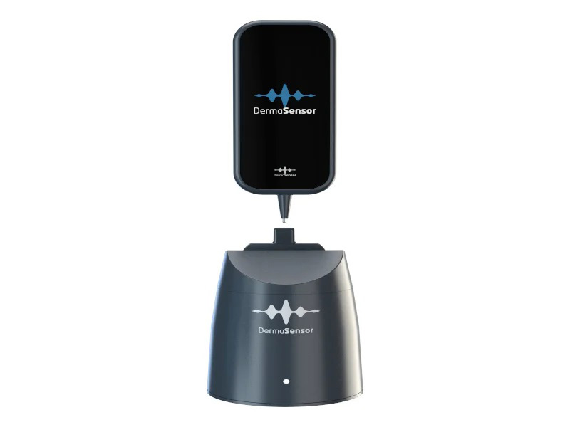
When it was first developed, ESS technology was the size of a microwave oven. Today, thanks to a decade of our team’s engineering advancement, you can hold it in the palm of your hand.
Harnessing the Power of AI
Even with cutting-edge ESS technology, there was no way to assess lesions objectively and efficiently. That’s why we developed an algorithm using machine learning. To build it, we collected thousands of dermatopathology-confirmed malignant and benign tissue samples. Then we compared the spectral data to the pathology report, testing and fine-tuning the algorithm in a process that continues today, as we constantly indentify and analyse new data, providing ongoing updates to our DermaSensor users.
Today, thanks to our proprietary DermaSensor algorithm, our device processes spectral data in a matter of seconds, rather than waiting days for a physician or labratory to return results.
Technology
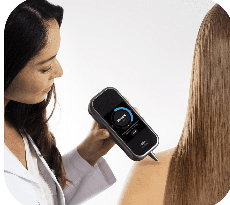
DermaSensor tip reflects and records quick bursts of light off the lesion’s cellular and sub-cellular content.
The light is analyzed by the built-in computer to provide information to help physicians assess skin lesions (including melanomas, squamous cell carcinomas, and basal cell carcinomas) to aid in a referral decision.
By examining the difference in light scattering, DermaSensor determines if the skin lesion is “Investigate Further” or “Monitor,” an immediate output.
Elastic Scattering Spectroscopy (ESS) has been validated in 30+ publications on clinical studies. Many studies have shown ESS to provide information that is comparable to histopathologic assessment in the analysis of cellular microscopic structure.
About ESS
DermaSensor uses Elastic Scattering Spectroscopy (ESS), a process which evaluates how photons scatter when reflected off of different cellular structures. Malignant lesions have been reported to have different cellular and sub-cellular structures than benign lesions, scattering light differently.