The Food and Drug Administration (FDA) has granted dual approvals for pegulicianine (Lumisight, Lumicell), an optical imaging agent, and Lumicell Direct Visualization System (DVS), a fluorescence imaging device, that may facilitate enhanced detection of residual breast cancer after removal of the primary tumor during lumpectomy procedures.
Collectively known as the LumiSystem, Lumisight and Lumicell DVS provide clinicians with real-time visualization that may offer a significant advance over current intraoperative imaging tools employed during lumpectomy procedures, according to Barbara L. Smith, M.D., Ph.D., the director of the Breast Program at Massachusetts General Hospital and a professor of surgery at Harvard Medical School.
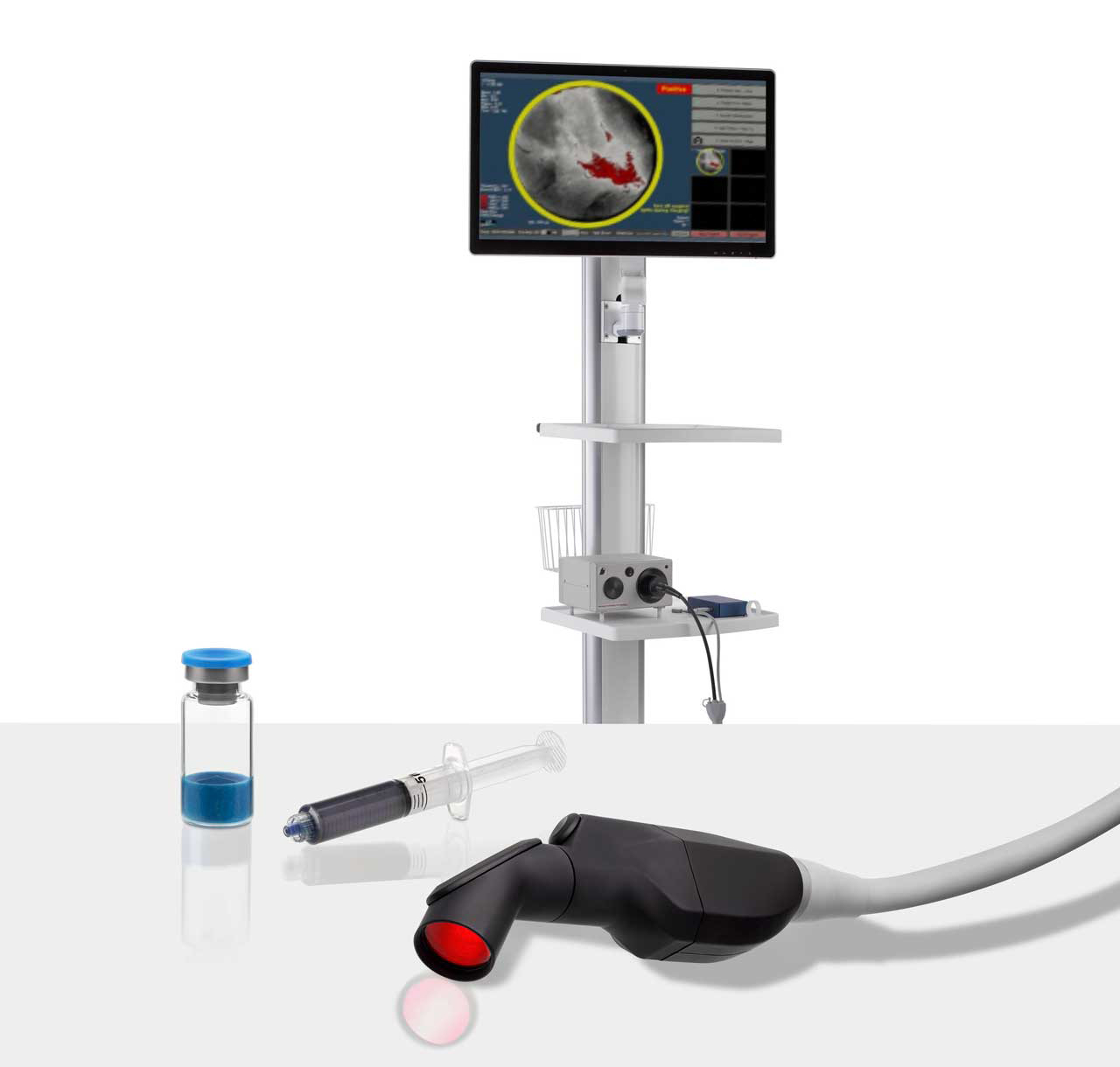
Lumicell’s approach starts with an injectable imaging agent, Lumisight (pegulicianine)—which is inactive at first but begins to glow when it interacts with particular enzymes found around tumor cells.
At the same time, the company’s computerized, handheld imaging probe scans the inside of the surgical cavity created during a breast-conserving lumpectomy and logs any signs of residual cancer hidden among the healthy cells.

“With LumiSystem, we will now have a technology that is clinically proven to achieve a more complete cancer resection during lumpectomy that could help some patients avoid a second surgery,” noted Dr. Smith.
Lumicell said the LumiSystem detects post-lumpectomy residual breast cancer with an 84 percent accuracy rate.1 In a 2023 study published in NEJM Evidence, Smith and colleagues noted that the LumiSystem enabled detection and removal of residual breast cancer after standard lumpectomy in 27 out of 357 patients with 22 of those patients originally deemed to have negative margins on standard margin assessments.
In the study, Dr. Smith noted unique attributes with the LumiSystem modality.
“pFGS (Pegulicianine fluorescence-guided surgery) is a cavity-based tool that identifies residual tumor within 2 to 5 mm from the surface of the lumpectomy cavity, rather than on the surface of the excised lumpectomy specimen, " wrote Dr. Smith and colleagues. "The pFGS cavity-based approach avoids the inherent problem of specimen-based approaches — correlating the location of tumor on an excised deformable specimen surface with the location of residual tumor in the breast cavity. pFGS also allows for repeat imaging of areas of concern during the initial operation to verify the removal of all positive signal areas."