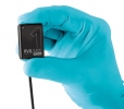
Digital dentistry can be defined as any dental technology or device that incorporates digital or computer-controlled components in contrast to that of mechanical or electrical alone. Several developments have been observed over the past few years that have enabled general dentistry to go to the next level. In this article, we’ll address a few such technologies.
Let’s take a look at some of the new technologies in the field of periodontology.
Diamond probe
A diamond probe combines manual probing with new technology. It’s plastic with black bands for visual scoring, and it measures the volatile sulfur compounds in the sulcus. The diamond probe offers more information than just measuring the sulcus depths. It informs the dentists of the sites that are showing signs of disease activity before the gingival attachment is lost and, at times, even before any bleeding is evident. It can also detect sulfides on the tongue.
Intraoral camera
In order to decide the nature of dental treatment and the right implementation, you need to have the exact images of the problem area in the teeth and their supporting parts. By using an intraoral camera, dentists can get a precise image, which helps in providing better dental care. This camera can also be used to educate patients about how they can improve their dental hygiene practices, like the areas they should focus on while brushing.
DIAGNOdent
This is a laser cavity detection technology that employs a laser diode to examine and sense early signs of tooth decay. You can get a digital print of even the smallest signs of decay. Since early detection means less complicated fillings, it helps in preserving more of the original tooth constitution. This technique is also safe as it uses light energy instead of X-rays.
T-Scan
This is a high-tech digital software that helps in recognizing premature contacts, interrelationship of occlusal surfaces and high forces, which ensures more accuracy and precision. Some benefits include:
-
Precise occlusal data and location of premature contacts
-
Reduced repeat visits to dentists
-
Enables the patient to understand better about occlusion
-
Preserve dental artistry by eliminating destructive forces
Bioprinting
You can “make” a tooth using Computer-Assisted Design/Computer-Assisted Manufacture (CAD/CAM) from a 3D scan. Research is being done to make recipes that could add tooth decay-fighting chemicals to 3D printed teeth. Some teams are engineering replacement for eroded jaw and gum tissue using the patient’s own cells. A team at Wake Forest Universityin North Carolina printed out human body parts from a mixture of live cells and gel laid down in layers to construct living human tissues. By doing this, they successfully "built" a jawbone.
Carbon-dioxide laser
This digital dentistry technology has removed the need of a scalpel due to its precision and ability to cut through both hard and soft tissues of the mouth. Carbon-dioxide lasers also allow for blood-free and anesthesia-free procedures, making dental treatments painless for the patients. It's great for patients with dental or pain anxiety as it is minimally invasive and highly effective. You can treat numerous conditions like fibroma removal, soft-tissue biopsies, soft-tissue crown lengthening and gum trimming.
Digital caries detection
Caries is a condition of decay and crumbling of the tooth or jaw bone. It's getting very hard to detect dentalcaries due to the large amount of fluoride that people consume in abundance, which in turns makes the tooth stronger. This technology helps dentists detect dental caries through the use of fluorescent lighting, which offers a reflection that allows the dentist to determine whether caries are present or not. They can also determine the severity of the caries once detected.
Cone beam CT
This pre-surgical imaging technique has made placements of dental implants extremely easy and predicable with a high success rate. The cone beam CT produces 3D images of the patient’s maxillofacial or oral anatomy. This 3D image is then used for preparing the implant surgical guides used by oral surgeons while fixing dental implants.
The future of dentistry is looking extremely bright. Waiting to adopt these areas of technology will leave you behind in the race. Determine which technology will enhance your practice, learn about it and make informed decisions before implementing.


