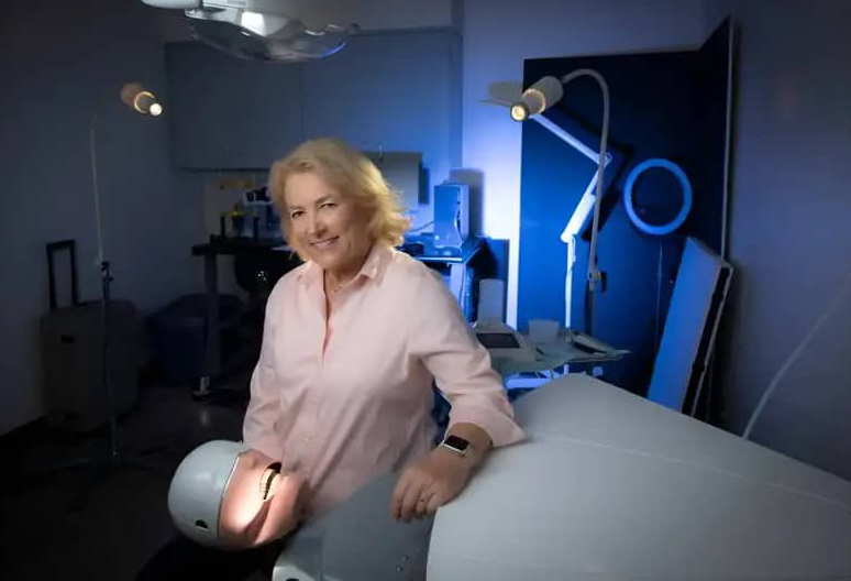
Oral cancer detection has always been challenging due to the varying appearance of lesions, often requiring a visual examination and biopsy. However, a new intraoral camera developed at the UCI Health Beckman Laser Institute & Medical Clinic promises to revolutionize this process. This cutting-edge device can identify cancerous lesions with up to 93% accuracy, a massive improvement from the standard 40-60% detection rates, offering hope for earlier and more effective treatment.
Intraoral cameras are essential tools in dentistry, shaped like pens and used to capture high-resolution images of teeth and oral tissues. These images are displayed on a screen for detailed assessment, and importantly, they pose no radiation risk to patients. The cancer-screening version of this device stands out for its ability to non-invasively detect suspicious areas, streamlining the diagnostic process.
An exciting feature of this innovation is the smartphone-compatible prototype designed as a phone case, making it easy to connect and visualize lesions. With its portability and user-friendly design, this intraoral camera could transform how oral cancer screenings are conducted, enhancing accessibility for healthcare professionals. Final testing and algorithm refinement are underway before mass production begins.
Dr. Petra Wilder-Smith, who led the development of this device, emphasizes the urgent need for better oral cancer outcomes. Despite advancements in treating other major cancers, oral cancer survival rates have worsened over time. Collaborating with the University of Arizona’s Wyant College of Optical Sciences, Dr. Wilder-Smith’s work focuses on reversing this trend through technological innovation.
The new intraoral camera has significant commercial and public health potential, with the promise of saving lives through earlier detection and intervention. As it moves closer to mass production, this breakthrough could redefine oral cancer care, setting a new standard for precision and efficiency in diagnosis.

