-
منوی اصلیبستن
-
Dental
-
-
-
Gloves
-
-
-
Gloves
-
-
-
Gloves
-
-
-
Gloves
-
-
-
Dental Finishing & Polishing
-
-
-
Gloves
-
-
-
Gloves
-
-
-
Gloves
-
-
-
Gloves
-
-
-
Gloves
-
-
-
Gloves
-
-
-
-
Gloves
-
-
-
Gloves
-
-
-
Gloves
-
-
-
-
-
Gloves
-
-
-
Gloves
-
-
-
Gloves
-
-
-
Gloves
-
-
-
Gloves
-
-
-
Gloves
-
-
-
Gloves
-
-
-
- Dandal Service
- Dandal Yar
- Mag
- Help
-
Corporate
-
- Login/ Register
-
منوی اصلیبستن
-
Dental
-
-
-
Gloves
-
-
-
Gloves
-
-
-
Gloves
-
-
-
Gloves
-
-
-
Dental Finishing & Polishing
-
-
-
Gloves
-
-
-
Gloves
-
-
-
Gloves
-
-
-
Gloves
-
-
-
Gloves
-
-
-
Gloves
-
-
-
-
Gloves
-
-
-
Gloves
-
-
-
Gloves
-
-
-
-
-
Gloves
-
-
-
Gloves
-
-
-
Gloves
-
-
-
Gloves
-
-
-
Gloves
-
-
-
Gloves
-
-
-
Gloves
-
-
-
- Dandal Service
- Dandal Yar
- Mag
- Help
-
Corporate
-
- Login/ Register
- Home
- Medical & Surgical Equipment
- Medical Equipment
- Ophthalmology
- Perimeter
- Haag Streit - Octopus 600 Perimeter
Haag Streit - Octopus 600 Perimeter - Dandal
-
In line with the demand of Dandal site customers for credit purchases, Dandal in cooperation with Saman Bank has provided a new service for dental and medical offices and treatment centers, which customers can use a credit card from the site at any time without any cash support.
- More Info
Haag Streit - Octopus 600 Perimeter
Out-of-StockFeatures:
- White-on-White Perimetry
- Glaucoma Screening Test
- Pusal Method
- Low Test-Retest Variability
- Fixation Control
- Out stock
- Free
Pay in installments
Choose your plan:
Current plan: 3Month
Description: 30+3
Down payment: تومان0 (25.00%)
Number of payments: 3
Tax: تومان0
Amount of payment: تومان0
Overpayment: تومان0
Total: تومان0
Current plan: Cash
Description: نقدی
Down payment: تومان0 (0.00%)
Number of payments: 1
Tax: تومان0
Amount of payment: تومان0
Overpayment: تومان0
Total: تومان0
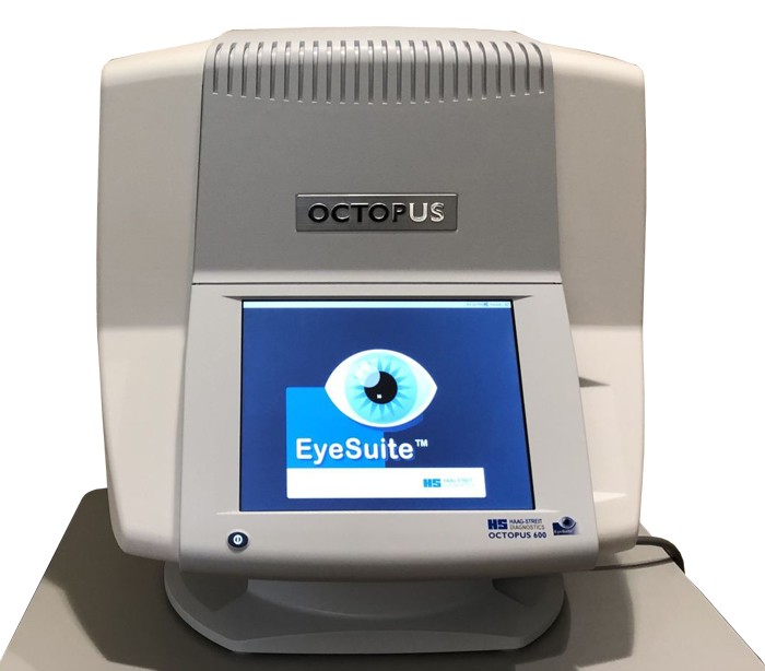
Octopus 600 Perimeter

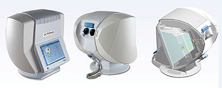
Detecting visual field loss at the earliest possible stage, defining the optimum treatment and following up the patient to decide on the necessity of treatment changes or surgery are the main tasks of every glaucoma specialist. Addressing these basic needs, Haag-Streit has introduced the Octopus 600 that combines early diagnosis and follow-up in a single compact-sized, standalone device with improved ergonomics both for operator and patient.
The device makes it possible to run standard central fields with minimum test duration, seamlessly integrated into your practice environment. This supports high patient throughput and effective practice management. With its built-in reliability features, the Octopus 600 is easy to use and delivers results you can trust.

Fast Screening
Distinguish between normal and abnormal visual fields in less than a minute with the novel Glaucoma Screening Test.
Central Field Standard
white-on-white perimetry
The Octopus 600 performs standard white-on-white threshold testing in just 2–4 minutes in the central visual field. A wide range of commonly used static test patterns, including G, 32, 30-2, 24-2, M, and 10-2 are also incorporated into the Octopus 600.
Perimetry Simplified
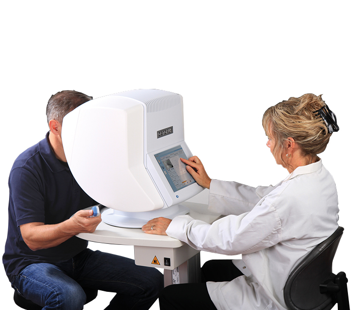
The compact Octopus 600 can be operated reliably outside of a darkroom and fits almost anywhere. With its improved usability and ergonomics, it truly simplifies perimetry both for the operator and the patient.
Reliable results made easy
Worry less about patient compliance. The Octopus 600 automatically recognises fixation losses and continously supports the correct patient and eye position for a reliable result you can trust.
OCTOPUS 600 Expert in Fast Screening and Follow-up
With the Octopus 600, Haag-Streit has introduced a new perimeter that combines both fast screening with the novel Pulsar stimulus and standard white-on-white perimetry for follow-up in one device. As a result, the instrument is capable of covering the most important stages of disease progression, allowing both for early diagnosis and follow-up. The Octopus 600 employs a new perimetry technology that makes use of a TFT-based monitor to produce stimuli and background. This allows the instrument to display complex stimuli such as Pulsar, which would otherwise not be possible with cupola-based or projection type perimeters.
Glaucoma Screening Test
Fast screening
Distinguish between normal and abnormal visual fields in less than a minute in healthy individuals. The test is purely qualitative and only distinguishes between normal and abnormal visual fields by presenting stimuli three times at a brightness that patients with normal vision should see. If never seen, then the visual field is flagged as abnormal with high reliability and the patient can be further tested. Because the test is efficient, it opens doors for more routine visual field testing to ensure no pathology goes undetected.
Pusal Method
The new pulse in perimetry
Pulsar is a patented1 flicker stimulus displaying a ring pattern especially developed for early glaucoma detection. The stimulus consists of two images, the phase and counterphase images that alternate with a frequency of 10 Hz over 500 ms. If flicker-sensitivity is reduced, the phase and counterphase images result in an overlapped image that is not visible anymore. By changing contrast and spatial resolution, full thresholds can be tested. Because the proven Pulsar method tests flicker and contrast sensitivity that are affected in early glaucoma, the method has been shown to be both sensitive and specific in the detection of glaucoma. This supports your correct diagnosis and allows you to start treatment in adequate time.
Low Test-Retest Variability
Made easy for patients
Unlike other early diagnostic methods that challenge patients with hard-to-distinguish answering criteria, Pulsar is easy for patients to answer: seen or not seen. Additionally, thanks to its design, it is relaxing to look at. This makes Pulsar a simple – even pleasant – test to take for patients. As a result, it shows low test-retest variability and only a minimal learning effect. These features make it a very suitable stimulus for screening purposes.
OCTOPUS 600
Central field standard white-on-white perimetry
The Octopus 600 offers a wide range of static test patterns, including 32 (which corresponds with 30-2), 24-2, and 10-2. In addition, there are two unique physiology-based patterns: the G-Program (a 30-degree field for glaucoma assessment) and the M-Program (a 10-degree field for analysing the macula). They are both correlated with a nerve fibre bundle map and thus make it possible to test the points which are most important for a structure-function correlation. These examination patterns offer a higher density of stimuli in the centre, which supports the discovery of paracentral scotomas that are often missed by the common 32 pattern. Combining the standard white-on-white perimetry with the Pulsar method for early glaucoma detection, the instrument is capable of covering the most important stages of disease progression, allowing for early diagnosis and follow-up. Being the first perimeter employing a flicker-free TFT monitor, stimuli can be soundlessly presented for increased reliability of the visual fields.
Top Fast-Thresholding Strategy
Increased Efficiency
Tendency Oriented Perimetry (TOP) presents a further optimisation in fastthreshold testing by reducing the examination time by nearly 80% to just 2–4 minutes7,8 compared to 6–8 minutes (Dynamic strategy) or 10–12 minutes (Normal strategy). The TOP algorithm is a systematic method which takes the correlation of the threshold values in neighbouring locations into account. Since the first test points are presented at a supra-threshold level, even inexperienced patients quickly understand the nature of the test.
Cluster Analysis
Providing meaningful results
Cluster analysis combines high sensitivity with good specificity9 . Test locations are grouped (clustered) along nerve fibre bundles, to better analyse changes in crucial areas such as the nasal step or the macula. This eliminates the time-consuming method of counting isolated points. A combined probability/deviation graph highlights pathological regions.
Polar Analysis
Combining structure and function
Combining the results of both structure and function (see picture: A) is key to obtaining a comprehensive assessment of the onset and progression of Glaucoma. With the Octopus Polar Analysis, the nerve fibre bundles that are in danger or defective are easily identified. Local defects are projected along the nerve fibres to the optic disk and are represented as red lines (B). The projected defects (C, D) are vertically mirrored and scaled with rings for 10, 20 and 30 dB deviation (E). The Octopus Polar Analysis allows for direct comparison with structural (F) findings10.
Fixation Control
Fixation losses due to low patient compliance are a major reason for unreliable visual fields. The Octopus 600 gives you less reason to worry about these. Blink Control, Pupil Position Control, Dart Control and Contact Control continously support the correct patient and eye position for a reliable result you can trust.
Product Details
Data sheet
- Company/ Brand
- Haag Streit
- Country
- Switzerland
- Shipping Weight
- 26 kg
- Display Screen
- TFT display
بیشتر
Data sheet
- Company/ Brand
- Haag Streit
- Country
- Switzerland
- Shipping Weight
- 26 kg
- Display Screen
- TFT display


